Extreme axial length in high myopia
Purpose. This case report describes a high axial myopia in a 71-year old female patient.
Material and Methods. Axial length was measured with the Myopia Master (Oculus Optikgeräte GmbH) and a fundus scan was performed with the EasyScan (Epitop GmbH).
Results. Axial length was 36.63 mm and 35.34 mm in the right and left eye. Both fundi presented myopic maculopathy with flecked chorioretinal atrophy and peripapillary atrophy. Visual acuity with contact lenses was 20/63 in the right eye and 20/50 in the left eye.
Conclusion. In the literature case reports of patients with extreme axial lengths were only found in patients of older age. High myopias can present with staphylomas, which leads to an increase in axial length. Therefore a screening of axial length could also be indicated in elderly, highly myopic patients.
Introduction
An axial length greater than 26 mm is associated with a 25 % increased risk of having a visual acuity of less than 20/40 at the age of 75 years. With an axial length exceeding 30 mm, the risk increases to 90 %.1 Which numbers can axial length reach in extreme cases? The following case describes a female patient with an extremely high ocular axial length.
Material and Methods
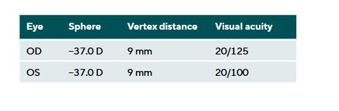
The 71-year-old female patient asked for a new contact lens fitting with RGP contact lenses. The results from the subjective refraction are shown in Table 1.
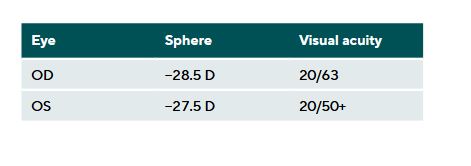
Rigid gas permeable contact lenses (Ascon, Hecht Contactlinsen GmbH) were fitted. Due to the increased retinal image size, visual acuity was improved compared to correction with glasses. (Table 2)
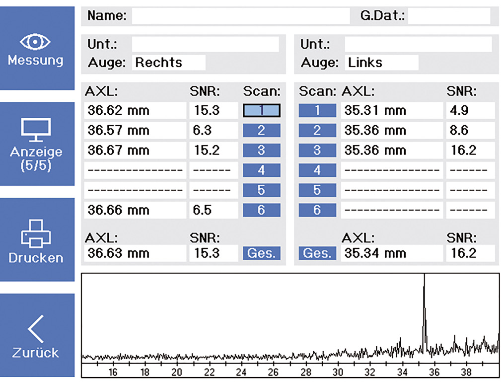
As part of the contact lens fitting, axial length measurements were taken using the Myopia Master (Oculus Optikgeräte GmbH). The axial length was 36.63 mm in the right eye and 35.34 mm in the left eye. Figure 1 shows the measurement results of the axial length measurement.
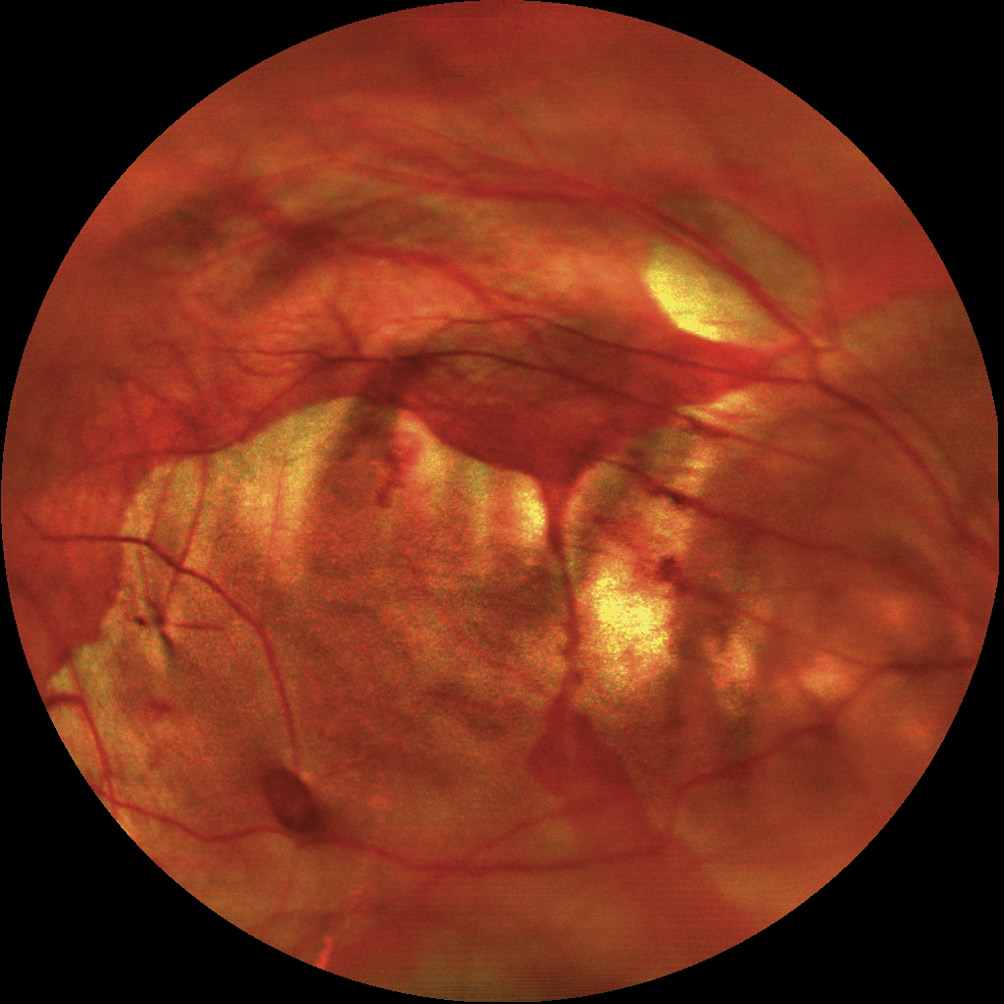
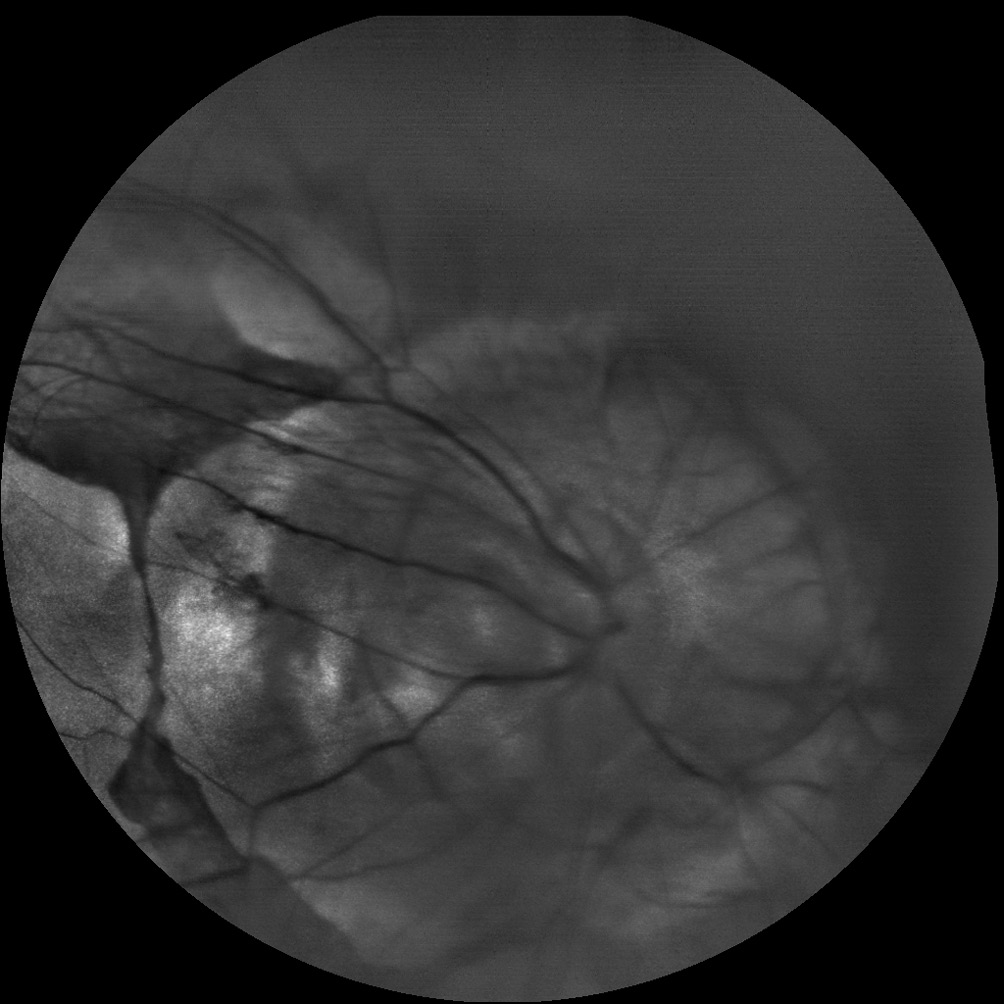
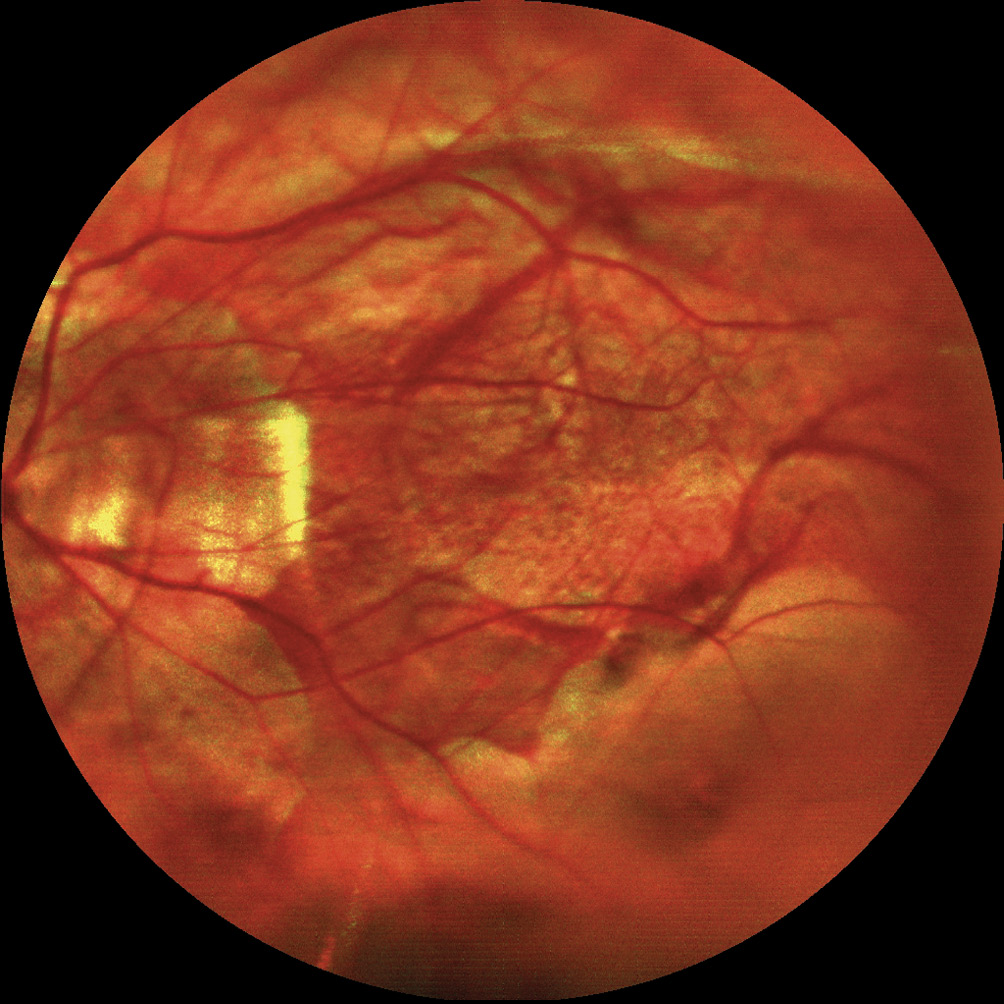
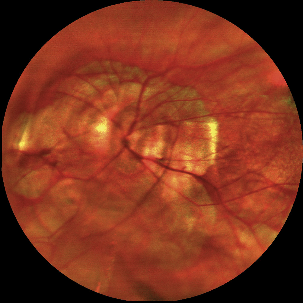
Additionally, retinal scans were taken using the EasyScan Fundus Scanner (Epitop GmbH). The patient had problems with central fixation in the right eye. Figure 2 shows the central retina of the patient with large atrophic areas and a Fuchs spot. There is also marked stretching of the blood vessels. Only the scan with the green laser showed a clear image of the papilla of the right eye (Figure 3). Therefore Figure 3 is presented as a black and white image. The right eye shows pronounced peripapillary atrophy. For the left eye (Figure 4 and Figure 5), the atrophic retinal areas spared the foveal region. Stretched blood vessels and peripapillary atrophy can be seen as well.
Discussion
The retina of the patient shows myopic maculopathy. Myopic maculopathy refers to typical myopic retinal changes, including diffuse or flecked chorioretinal atrophy, myopic macular degeneration, lacquer cracks, Fuchs spots, myopic choroidal neovascularization, posterior staphylomas and peripapillary atrophy. The risk of these changes increases with an axial length of 25.9 mm in men and 25.3 mm in women.2 A staphyloma, defined as a bulging of the sclera with a radius smaller than the surrounding scleral radii, causes further elongation of the eye. However, only macular staphylomas can be measured in axial length measurements, which occur more frequently than papillary or nasal staphylomas.3 Women are more frequently affected by ocular changes due to staphylomas. As the patient ages, staphylomas can change more rapidly. Therefore, in highly myopic patients changes in axial length can still be observed at an advanced age.4 Furthermore, Du et al. showed that in highly myopic individuals, axial lengths continue to increase throughout life, regardless of staphyloma formation.5 However, Gaucher et al. reported cases where the axial length in highly myopic patients decreased due to dome-shaped bulging of the sclera in the macular region. This causes an inward indentation of the macula, which can be detected on OCT. In these cases a thickening of the subfoveal sclera can be observed. Dome-shaped maculae are typically associated with atrophic changes in the retinal pigment epithelium and subretinal fluid.6
In population studies, axial lengths over 35 mm are rare. In Russia, axial lengths were measured via ultrasound in 5707 subjects between 40 and 94 years of age, with values ranging between 19.02 mm and 32.87 mm.7 A larger study from Japan measured axial lengths in 22,379 subjects using the coherence interferometer OA-1000 (Tomey, Japan), with only four eyes showing axial lengths exceeding 35 mm. All participants with these extreme axial lengths were over 60 years old. The axial length distribution of the studies showed an increase of numbers of very high axial lengths with advancing age.8 Therefore, it can be assumed that such extreme axial lengths develop later in life, often due to the formation of staphylomas. Two additional case reports were found with even longer axial lengths.
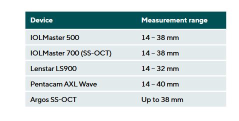
Vasallo and Fenech described in 2020 the longest axial length at the time, 38.34 mm in the right eye and 38.31 mm in the left eye in a 47-year-old patient of undefined gender. These measurements were quantified using ultrasound, because measurements with the Lenstar LS900 (Haag-Streit) were not possible.9 In 2024, an even longer eye was described in a 53-year-old woman, with axial lengths of 40.59 mm in the right eye and 40.84 mm in the left eye, measured with the Argos SS-OCT (Alcon). Axial length measurements in this patient were taken at different times with various devices. At the age of 41, the same patient’s axial lengths were measured using ultrasound, showing 40.59 mm in the right eye and 38.29 mm in the left eye. Measurements with the
IOLMaster 500 (Carl Zeiss Meditec AG), IOLMaster 700 (Carl Zeiss Meditec AG), and Pentacam AXL-Wave (Oculus Optikgeräte GmbH) failed at their respective device-specific maximum values (due to the length of the reference-arm). The authors noted that the measurement with the Argos SS-OCT was possible, despite the official measurement range supports only measurements up to 38 mm.10
Conclusion
Axial lengths greater than 35 mm are rarely measured. Depending on the measuring device, the technical limits for axial length measurements range between 32 mm and 40 mm. Longer axial lengths cannot be measured due to the design limitations of the reference arm in biometric devices.
This case presents the longest axial length measurement to date with the Myopia Master. Since no other biometer or ultrasound device was available, the accuracy of the measured axial length could not be confirmed by an independent measurement. The case shows that measurements of axial lengths over 36 mm are possible with this device. According to the data sheet, the Myopia Master allows axial length measurements of up to 40 mm.
Because in highly myopic patients, axial length can change even at advanced age due to staphylomas (axial elongation) or the appearance of a dome-shaped macula (axial shortening), axial length measurements can also be integrated into optometric examinations for this group of patients.
Conflict of interest
The author declares that she has received lecture honoraria from the company Oculus Optikgeräte GmbH within the last three years.

About Carolin Truckenbrod
Dr. rer. med., M.Sc., Dipl.-Ing. (FH) - Truckenbrod Augenoptik, Leipzig, Germany, LIFE Leipzig Research Center for Civilization Diseases, Universität Leipzig, Leipzig, Germany
and Myopic Maculopathy: The Hisayama Study. Ophthalmol. Retina.,
3, 867–873.
Increase of Axial Length and Its Risk Factors in Adults With High Myopia. JAMA Ophthalmol., 39, 1096–1103.
Arslangareeva, I. I., Salavatova, V. F., Bikbova, G. M., Panda-Jonas, S.,
Nikitin, N. A., Zaynetdinov, A. F., Nuriev, I. F., Khikmatullin, R. I., Uzian-
baeva, Y. V., Yakupova, D. F., Aminev, S. K., Jonas, J. B. (2019). Axial
length and its associations in a Russian population: The Ural Eye and Medical Study. PloS One. 14, e0211186.
Tsuchiya, N., Nakamura, T., Ishikuro, M., Obara, T., Miyazawa, A., Homma, K.,
Ido, K., Taira, M., Kobayashi, T., Shimizu, R., Uruno, A., Kodama, E. N.,
Suzuki, K., Hamanaka, Y., Tomita, H., Sugawara, J., Suzuki, Y., Nagami, F.,
Ogishima, S., Katsuoka, F., Minegishi, N., Hozawa, A., Kuriyama, S., Yaegashi, N., Kure, S., Kinoshita, K., Yamamoto, M. (2022). Genome-wide Association Study of Axial Length in Population-based Cohorts in Japan: The Tohoku Medical Megabank Organization Eye Study. Ophthalmol. Sci. 2, 100113.