Facial Nerve Palsy: Causes, Ocular Manifestations, and Treatment Options
Purpose. The aim of this study is to describe the causes, clinical manifestations, and treatment options for peripheral facial palsy, with a particular focus on ocular complications.
Material and Methods. This is a narrative review based on current literature and clinical experience. Etiological factors, functional limitations, and therapeutic approaches are systematically summarized and supplemented by a case study on scleral lens management.
Results. Facial nerve palsy is predominantly idiopathic (Bell’s palsy), but can also be caused by viral, systemic, traumatic, or neoplastic factors. Clinically, it leads to facial asymmetry, functional limitations in speaking and eating, and psychosocial distress. Ocularly, lagophthalmos is the primary concern, which can lead to impaired tear film distribution, keratopathies, and potentially severe corneal damage. Additionally, hearing impairments and balance disorders may occur. A multidisciplinary approach is required for treatment. In addition to conservative measures (moisturization, protection, functional therapy), scleral lenses demonstrate effective improvement of the ocular surface and visual function.
Conclusion. Peripheral facial palsy is a complex condition with potentially serious ocular consequences. Early diagnosis and personalized, interdisciplinary treatment are crucial for preventing irreversible damage and sustainably improving the quality of life for those affected.
Introduction
Peripheral facial nerve palsy is a disorder of the seventh cranial nerve, the facial nerve, affecting its peripheral course after it leaves its nucleus, which leads to paralysis and, in the long term, to abnormal control of the facial muscles in some cases. This paralysis usually occurs on one side, but can also occur on both sides and may have a variety of causes. This paper describes the causes of facial nerve palsy as well as its effects on the body, particularly on the eye. Finally, the various prognoses for facial nerve palsy are discussed.
Material und Methoden
Der klinische Artikel basiert auf einer zusammenfassenden Literaturübersicht zu Gesichtsnervenlähmungen, deren Ursachen, okulären Manifestationen und therapeutischen Behandlungsmöglichkeiten. Ergänzt wird dies durch die klinischen Erfahrungen der Autoren. Der aktuelle Wissensstand hinsichtlich der Ätiologie, der damit verbundenen Funktionsbeeinträchtigungen und der Behandlungsansätze wird systematisch zusammengefasst. Darüber hinaus wird die Behandlung mit Sklerallinsen anhand eines repräsentativen klinischen Fallbeispiels beschrieben.
Causes of Facial Nerve Palsy
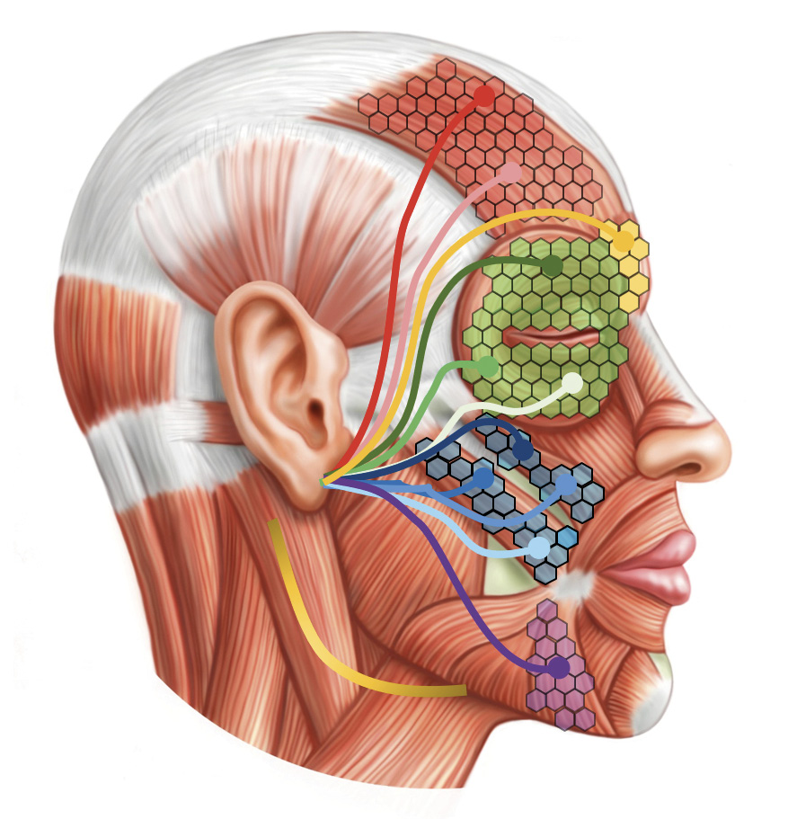
Facial nerve palsy can result from a wide range of underlying conditions. However, despite extensive diagnostic evaluation, no identifiable cause is found in the majority of cases.1 Such cases are classified as idiopathic facial nerve palsy, commonly referred to as Bell’s palsy.2 The condition typically develops within minutes to a few hours and, by definition, occurs without an identifiable cause.3
Viral infections may be detected by serological testing or cerebrospinal fluid analysis.4 In addition, characteristic skin lesions, particularly around the ear, may provide important diagnostic clues. Even in apparently idiopathic cases, however, a viral etiology is frequently suspected, most commonly involving herpes simplex virus (HSV). Reactivation of HSV may induce inflammation of the facial nerve, resulting in nerve dysfunction and subsequent facial paralysis.
Another important viral cause is varicella-zoster virus (VZV), the causative agent of chickenpox and herpes zoster. Reactivation of VZV may lead to Ramsay Hunt syndrome, in which the virus affects the facial nerve, causing facial nerve palsy. The associated inflammation and nerve edema can result in temporary impairment of facial nerve function and, in some cases, permanent neurological deficits.5,6
Systemic diseases such as diabetes mellitus, multiple sclerosis (MS), and Guillain-Barré syndrome (GBS) are well-recognised causes of facial nerve palsy.7 In diabetes mellitus, poor glycaemic control may result in microangiopathic damage to the blood vessels supplying the facial nerve, leading to impaired perfusion, ischaemia, and subsequent nerve dysfunction. In MS, an autoimmune demyelinating disease of the central nervous system, immune-mediated destruction of the myelin sheath may involve the facial nerve, impairing nerve conduction and function. The resulting inflammation and demyelination may ultimately cause facial paralysis. Guillain-Barré syndrome is an acute autoimmune neuropathy that typically develops following an infection.7 In GBS, immune-mediated inflammation of the peripheral nerves may also affect the facial nerve, resulting in temporary facial paralysis.
Additional infectious causes include Borrelia burgdorferi, human immunodeficiency virus (HIV), poliovirus, mumps virus, and Epstein–Barr virus, the causative agent of infectious mononucleosis.8 Tuberculous otitis media has also been reported as a rare cause of facial nerve palsy.9 Traumatic aetiologies include traumatic brain injury and surgical procedures involving the head and neck region.10 In addition, cerebrovascular events affecting the facial motor pathway may also present with facial weakness or paralysis.11
Finally, facial nerve palsy may also result from neoplastic processes. These include schwannomas involving the vestibulocochlear nerve or the facial nerve,12 as well as malignant neoplasms of the parotid gland, such as adenoid cystic carcinoma.13 In addition, metastatic involvement of the facial nerve may occur in association with primary malignancies of the orbit, lung, breast, or kidney, as well as locally invasive cutaneous malignancies, including infiltrative basal cell carcinoma. (Figure 1)
Effects of Facial Nerve Palsy
Facial Expression Disorder
Facial nerve palsy has profound effects on the entire face, particularly the eye.14 As the facial nerve provides motor innervation to the muscles of facial expression, paralysis results in facial asymmetry and impaired facial function. The loss of muscle activity compromises facial expression and the coordinated execution of facial movements.
One of the most characteristic clinical features is unilateral facial asymmetry. On the affected side, the facial muscles are either completely paralysed or exhibit markedly reduced function, resulting in the typical appearance of unilateral facial paralysis. This asymmetry becomes particularly evident during voluntary facial movements such as smiling, blinking, or frowning, when the unaffected muscles contract normally while those on the affected side remain inactive.
The corner of the mouth is often displaced toward the unaffected side, resulting in a characteristic unilateral facial droop and marked facial asymmetry. Consequently, the ability to express emotions through facial expressions is substantially impaired. Dysfunction of the orbicularis oculi muscle, which is responsible for blinking and eyelid closure, is among the most clinically significant manifestations of facial nerve palsy. Incomplete eyelid closure (lagophthalmos) increases tear film instability and ocular surface exposure, predisposing patients to dry eye disease, exposure keratopathy, corneal epithelial defects, and secondary infections. Likewise, smiling and showing the teeth become asymmetrical because the perioral muscles on the affected side are unable to contract normally.
The severity and distribution of facial nerve palsy vary considerably. Paralysis may affect the entire hemiface or be limited to specific regions, including the forehead, the periocular region, or the perioral musculature.
Difficulties with Speech and Eating
Facial nerve palsy may also impair speech and oral motor function. Weakness of the perioral musculature compromises lip movement and articulation, frequently resulting in dysarthria.15 In addition, impaired function of the orbicularis oris muscle reduces lip closure, leading to difficulties with chewing, drinking, and the controlled oral transport of food, which may increase the risk of oral injury and dental complications.16 These functional impairments are often among the earliest symptoms reported by patients. Inadequate lip seal may also result in involuntary drooling (sialorrhoea) on the affected side, a characteristic clinical feature of facial nerve palsy.
Psychosocial Effects
The consequences of facial nerve palsy extend beyond physical impairment and often have a substantial psychosocial impact.17 Difficulties with speech may compromise verbal communication, while reduced facial expressiveness impairs nonverbal communication, thereby affecting social interaction and interpersonal relationships. Many patients report diminished self-esteem and social withdrawal, as the visible facial asymmetry is frequently misunderstood and may result in discomfort, embarrassment, or social stigmatization. The reduced ability to convey emotions through facial expressions can further contribute to frustration and emotional distress. Psychological sequelae are common and encompass a broad spectrum of conditions, including anxiety and depression, particularly in patients at risk of vision loss.18
Effects of Facial Nerve Palsy on Hearing
Although facial nerve palsy primarily affects facial motor function, it may also be associated with auditory symptoms due to the close anatomical relationship between the facial nerve (cranial nerve VII), the vestibulocochlear nerve (cranial nerve VIII), and the structures of the middle and inner ear. One of the most characteristic auditory manifestations is hyperacusis, defined as increased sensitivity to ordinary sound levels.
The facial nerve provides motor innervation to the stapedius muscle, a small middle ear muscle that regulates stapes movement and contributes to the attenuation of loud sounds. Facial nerve paralysis results in impaired stapedius muscle function, reducing this protective mechanism and potentially causing hyperacusis. Consequently, everyday sounds may be perceived as excessively loud or uncomfortable by affected individuals.19 Tinnitus, described as the perception of ringing, buzzing, or other sounds in the absence of an external acoustic stimulus, may also occur in association with facial nerve palsy. Altered facial nerve function and abnormal activation of related muscular structures may contribute to subjective auditory sensations, particularly during facial movements.
In some cases, facial nerve palsy may occur in combination with temporary or permanent hearing impairment. This may result from concurrent involvement of the vestibulocochlear nerve or inflammatory processes affecting the inner ear. A well-known example is Ramsay Hunt syndrome, in which reactivation of varicella-zoster virus (VZV) affects both the facial nerve and the vestibulocochlear nerve, potentially resulting in the combination of facial nerve palsy and sensorineural hearing loss.
Effects on the Eye
Blinking plays a crucial role in maintaining ocular surface integrity by distributing the tear film and providing continuous lubrication and protection of the eye. In facial nerve palsy, impaired function of the orbicularis oculi muscle results in reduced or incomplete eyelid closure, leading to lagophthalmos.20
Incomplete eyelid closure compromises tear film distribution and increases ocular surface exposure, particularly affecting corneal integrity. Insufficient protection of the corneal surface may result in tear film instability, localized desiccation, and epithelial microdefects, clinically referred to as superficial punctate keratitis. These epithelial disruptions may facilitate microbial invasion and increase the risk of infectious keratitis.
Patients may experience a range of visual symptoms, including fluctuating or blurred vision, reduced visual acuity, increased glare sensitivity, and ocular discomfort or pain. In severe cases, microbial keratitis can lead to corneal scarring, potentially resulting in permanent visual impairment in the affected eye.
Impaired tear film distribution, combined with reduced blinking, may also affect the conjunctiva and contribute to ocular surface inflammation. Clinical manifestations include conjunctival hyperaemia, chemosis, and increased tear production (epiphora). Persistent exposure and desiccation of the ocular surface may further progress to corneal epithelial breakdown and, in severe cases, corneal ulceration.
In addition to lagophthalmos, which represents the most common eyelid abnormality associated with facial nerve palsy, other eyelid malpositions may occur, including upper eyelid ptosis, entropion, and ectropion. Particularly the latter two conditions, when combined with incomplete eyelid closure, can further compromise ocular surface protection and increase tear film evaporation. Additional corneal damage may result from mechanical trauma caused by misdirected eyelashes (trichiasis) rubbing against the corneal surface.
Facial nerve palsy may also be associated with altered periocular sensation. Increased sensitivity to external stimuli, such as light, wind, or dust, may occur and is referred to as hyperaesthesia. Conversely, reduced sensory perception (hypoaesthesia) may impair the patient‘s ability to detect ocular irritation or injury at an early stage.21
These functional impairments highlight the need for comprehensive management addressing both the medical and psychosocial consequences of facial nerve palsy. Such an approach aims to improve patients’ quality of life and support their participation in social interactions.22 Particularly with respect to ocular involvement, the potential risk of vision loss may lead to significant restrictions in daily activities, including professional and social participation.
Prognosis
The prognosis for facial nerve palsy varies depending on the cause, severity, and timing of treatment.23,5 In cases of idiopathic Bell’s palsy, most patients make a full recovery within weeks to months.24 However, some patients experience only partial improvement in their symptoms, which can lead to permanent functional impairment. In these cases, the axons the individual nerve fibers that carry signals to the muscle fibers have often been so severely damaged, for example by trauma or inflammation, that they have died at the site of the injury. Fortunately, peripheral nerves – which include the cranial nerves have the ability to regenerate. At the site of injury, several new nerve sprouts often form, attempting to reconnect with the muscles they previously innervated.
This regenerative process typically requires three to six months; therefore, in cases of extensive axonal injury, functional recovery may not become clinically apparent until after this period. During reinnervation, however, aberrant nerve regeneration may occur, resulting in misdirected connections between nerve fibres and target muscles. A characteristic example is involuntary eyelid closure during movements of the mouth, such as lip pursing. This phenomenon is referred to as oro-ocular synkinesis.
In such cases, the initially flaccid facial paralysis observed during the acute phase may progress to chronic synkinetic facial palsy, which can result in persistent functional limitations. Although these conditions are often still described as facial palsy, the affected muscles are usually reinnervated and retain motor function; however, their activation patterns are abnormal and poorly coordinated. Consequently, facial expressions remain impaired despite preserved
muscle activity.
Only a small proportion of patients, particularly those with extensive or long-segment nerve injuries, develop persistent flaccid paralysis of the facial muscles. For patients in the acute phase, the likelihood of recovery is a major concern, highlighting the clinical importance of reliable prognostic indicators.
Several factors influence the prognosis of facial nerve palsy. A milder initial presentation is generally associated with a more favourable outcome compared with complete paralysis. Younger age has also been identified as a positive prognostic factor. Early diagnosis and timely initiation of appropriate treatment may further improve recovery prospects. The underlying aetiology remains one of the most important determinants of outcome. For example, patients with idiopathic facial nerve palsy (Bell’s palsy) generally have a more favourable prognosis than those with tumour-associated facial nerve palsy.
Treatment and Management
The treatment and management of ocular complications associated with peripheral facial nerve palsy are of paramount importance to prevent ocular surface damage and preserve patients’ quality of life. As incomplete eyelid closure represents one of the most significant consequences of facial nerve dysfunction, targeted therapeutic strategies are required to protect the ocular surface and maintain adequate corneal lubrication.
This section outlines current treatment approaches and protective measures, including lubrication therapy, eyelid management strategies, and the use of scleral lenses as a therapeutic option for patients with persistent ocular surface exposure.
Specialized Facial Therapy for Incomplete Eyelid Closure
Incomplete eyelid closure may occur even in individuals without facial nerve palsy; however, the severity is generally limited and does not exceed grade 2. In contrast, patients with facial nerve palsy typically present with more pronounced eyelid closure deficits, corresponding to grades 3 and 4. (Figure 2)
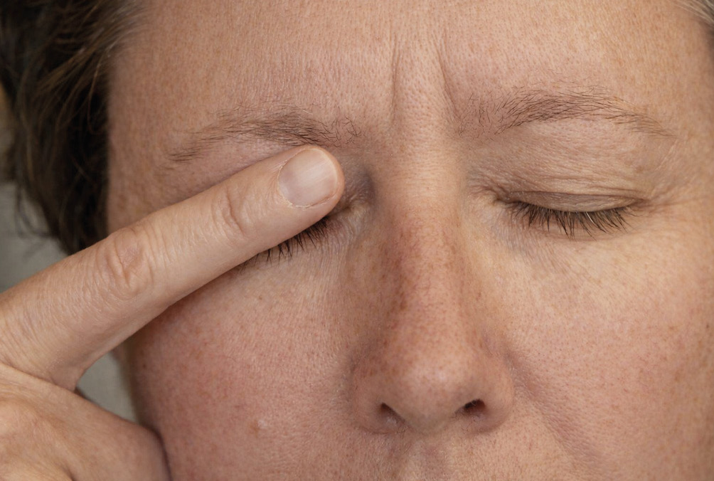
In German-speaking countries, the management of peripheral facial nerve palsy often involves physical therapists or speech and language therapists with specialized additional training. A central component of therapy is educating patients on effective strategies to protect the ocular surface despite incomplete eyelid closure. For example, the application of lubricating eye drops and ointments requires adaptation, as reduced or absent blinking impairs their uniform distribution across the ocular surface. Therefore, manual assistance, such as deliberate eyelid closure or gentle spreading of the medication, may be necessary to ensure adequate ocular surface coverage. (Figure 3)

Eye Protection at Night
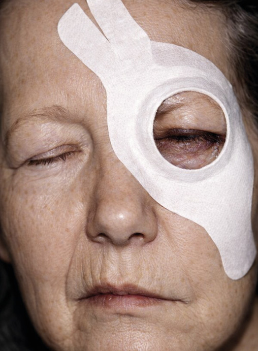
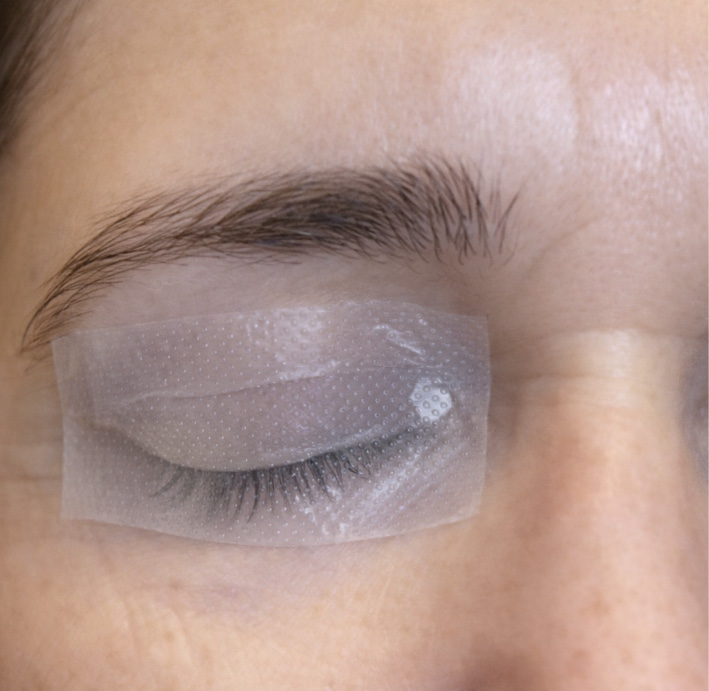
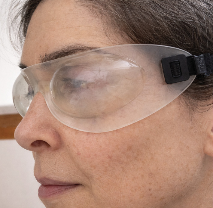
Nocturnal eye protection represents a particular challenge in patients with facial nerve palsy, as reduced blinking during sleep combined with incomplete eyelid closure may significantly increase ocular surface exposure and tear film evaporation. During the acute phase, conventional management commonly includes the application of lubricating ophthalmic ointment together with mechanical eyelid closure using an eye patch or the use of moisture chamber goggles.
However, these approaches require individual adaptation, as patient tolerance varies and some individuals may experience difficulties with ointment application, adhesive materials, or maintaining adequate eyelid coverage throughout the night. Moisture chamber goggles, such as the EyeSeal 2.0, provide a humidified microenvironment around the eye and may help reduce tear evaporation and ocular surface desiccation during sleep. Their effectiveness, however, depends on stable positioning and patient compliance; in individuals with restless sleep or frequent nocturnal movement, displacement of the goggles may limit their protective benefit. (Figure 4, Figure 5, Figure 6)
Eye protection during the day
During daytime, continuous use of a bandage glass or adhesive eye shield is rarely required. Instead, the focus should be on measures aimed at protecting the ocular surface from environmental stressors such as wind, sunlight, and air drafts. These measures include the use of protective eyewear or hats, as well as regular manual stretching of the upper eyelid to prevent progressive shortening and support adequate corneal lubrication. Such interventions may be instructed and supervised by a specialized therapist and should be individually adapted to the patient’s functional status and specific needs.
Treatment by the multidisciplinary team
The management of patients with peripheral facial nerve palsy often requires a multidisciplinary approach involving ophthalmologists, optometrists, and facial therapists. Close collaboration between these specialists is essential to ensure comprehensive ocular care and to minimize the risk of ocular surface complications. Treatment decisions should consider individual patient factors, including age, the severity of lagophthalmos, and the anticipated duration until restoration of eyelid closure. Therapeutic interventions should be continuously adapted according to the course of recovery to protect the cornea and prevent irreversible ocular surface damage.
Case Report: Scleral Lens Fitting in Unilateral Facial Nerve Palsy
Particular attention should be given to scleral lens fitting, as it represents an effective therapeutic option for the management of lagophthalmos and associated ocular surface disease. Scleral lenses completely vault the cornea and maintain a fluid reservoir over the ocular surface, thereby providing continuous hydration and mechanical protection against desiccation.
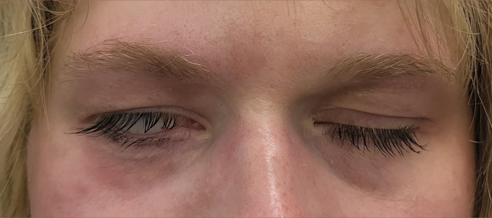
The following case report describes a patient with unilateral facial nerve palsy. The 30-year-old patient developed chronic facial nerve palsy during infancy following forceps-assisted delivery. (Figure 7) At the age of 10 years, she underwent a dynamic muscle transfer using leg muscle tissue to improve facial function. As part of her conservative ocular management, she requires frequent application of lubricating eye drops, the use of soft bandage contact lenses, and nocturnal application of ointment combined with bandage glass protection.
However, the soft bandage contact lens is frequently lost, which is likely related to incomplete eyelid closure and upward displacement of the lens caused by Bell’s phenomenon.
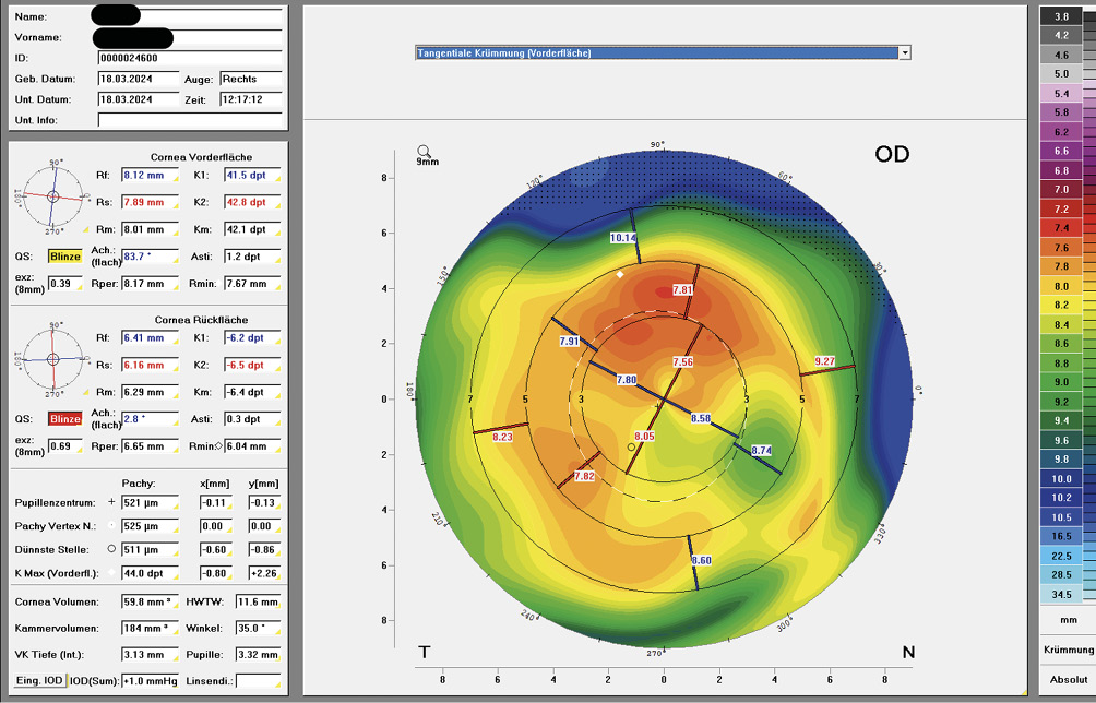
The best-corrected visual acuity (BCVA) prior to scleral lens fitting was 20/100 in the right eye (OD) and 20/16 in the left eye (OS). Corneal topography of the right eye revealed an irregular pattern, which was likely associated with chronic ocular surface changes resulting from longstanding lagophthalmos and exposure-related dryness. (Figure 8)
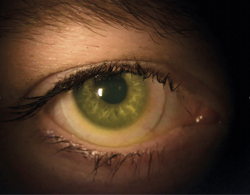
Following successful scleral lens fitting, the patient reported a marked improvement in ocular symptoms and overall comfort. (Figure 9) She wears the scleral lens daily for approximately 10–12 hours and describes a significant improvement in her quality of life. Best-corrected visual acuity in the right eye (OD) improved to 1.0, contributing to functional binocular vision.
It should be noted that, despite the continuous fluid reservoir provided by the scleral lens, surface drying of the lens may occur during wear. In such cases, rewetting can be achieved by manually spreading the tear film across the lens surface through deliberate blinking or gentle eyelid manipulation. The same principle applies when applying artificial tears, which need to be distributed evenly over the anterior lens surface to restore optical clarity and comfort.
Additional medical treatments
In addition to the therapeutic approaches described above, various medical and surgical options are available for the management of peripheral facial nerve palsy and its associated ocular complications. Surgical interventions include tarsorrhaphy, a procedure in which the palpebral fissure is partially or completely narrowed by joining the eyelids to improve ocular surface protection. This approach may be considered in severe cases of lagophthalmos when conservative measures fail to provide adequate corneal protection.
However, with the availability of effective conservative treatment strategies and alternative surgical procedures that better preserve eyelid function, tarsorrhaphy is now rarely required. Another surgical option is the implantation of upper eyelid weights, which facilitate eyelid closure by utilizing gravity-assisted descent of the upper eyelid. This approach can be particularly effective in upright positions, such as sitting or standing, where gravitational forces support eyelid closure.
Another therapeutic option is the use of botulinum toxin. Injection of botulinum toxin into the levator palpebrae superioris muscle can induce temporary pharmacological ptosis, thereby improving eyelid closure and providing protection of the ocular surface.25 This approach may be considered in cases of long-standing facial nerve palsy when conservative management alone does not provide sufficient ocular surface protection.
Dynamic muscle transfer techniques are recommended for patients with chronic facial nerve palsy persisting for more than 1.5 years, particularly when spontaneous recovery is unlikely. In cases of confirmed muscle atrophy, reconstructive procedures may include transfer of a segment of the temporalis muscle to the cheek or a free functional muscle transfer using, for example, a gracilis muscle graft harvested from the thigh.1,26
These therapeutic options provide additional strategies for preserving and improving ocular surface health in patients with peripheral facial nerve palsy, particularly when integrated into a comprehensive multidisciplinary treatment approach.
Conclusion
Facial nerve palsy is a complex condition with diverse etiologies and clinical manifestations. Ocular involvement, particularly due to lagophthalmos and impaired eyelid function, can lead to significant morbidity and requires early recognition and targeted management to prevent long-term ocular surface damage. Prognosis varies considerably and depends on multiple factors, including the underlying cause, severity of nerve impairment, and response to treatment.
The management of ocular complications associated with peripheral facial nerve palsy requires an individualized and multidisciplinary approach that is continuously adapted to the patient’s course of recovery. Specialized conservative treatment options, such as scleral lenses, provide effective means of protecting the ocular surface, maintaining visual function, and improving quality of life. Close collaboration among ENT specialists, ophthalmologists, optometrists, and facial therapists is essential to ensure comprehensive care tailored to the individual needs of each patient.
Conflict of Interests
The authors declare that there is no conflict of interests regarding the methods and devices mentioned in the article.