Scleral Lens Fitting in Keratoconus – Algorithms for Clinicians
Purpose. To present a structured, evidence-based clinical algorithm for scleral lens fitting in advanced keratoconus that integrates disease progression management, optical power optimization, and scleral asymmetry correction.
Material and Methods. This review synthesizes current evidence on scleral lens dynamics, keratoconus progression, high-powered lens management, and corneoscleral topography. The algorithm is built around three pillars: (1) a 100 micron progression safety margin, (2) strategic manipulation of back optic zone radius (BOZR) and diameter (BOZD) to optimize optical performance, and (3) management of scleral asymmetry using modern imaging. These concepts are integrated into distinct pathways for centered versus decentered ectasia.
Results. The algorithm provides a stepwise decision tree incorporating pre fitting assessments, foundational fit with central clearance of 200–300 µm (plus 100 µm for progressive disease), and over refraction to guide BOZR/BOZD manipulation for high powered lenses. Distinct pathways are outlined for centered ectasia (peripheral clearance management, oblate designs for high myopia with BOZD decreased for high myopia, and BOZR steepening with BOZD increase for high hyperopia) and decentered ectasia (optic zone decentration, quadrant specific modifications, and customized free form lenses). A troubleshooting protocol addresses common complications, and piggyback systems are discussed as a temporary rescue option.
Conclusion. Successful scleral lens fitting in advanced keratoconus requires a systematic, parameter driven approach that respects corneal and scleral topography, accounts for disease progression, and addresses refractive error through targeted BOZR/BOZD manipulation. The presented algorithm offers practitioners a structured framework to achieve reproducible outcomes and long term ocular health.
Introduction
Keratoconus is a progressive corneal ectatic disorder characterized by stromal thinning (most commonly in the inferotemporal region), corneal protrusion, and irregular astigmatism, leading to significant visual impairment.1,2,3,4 Traditionally considered noninflammatory,5,6 emerging evidence links it to altered inflammatory mediators, suggesting underlying ocular inflammation.7,8,9,10,11,12,13 Although bilateral, one eye is usually more severely affected.14,15,16,17,18 It affects all sexes and ethnicities, often as an isolated condition but sometimes with other ocular or systemic diseases.5
Keratoconus remains a leading indication for corneal transplantation, yet the vast majority of patients can be successfully rehabilitated with contact lenses.19,20 The evolution from corneal lenses to corneoscleral and fully scleral designs has broadened the range of available options for keratoconus management. Unlike corneal lenses that rest on the irregular ectatic surface – often inducing apical scarring or decentration 21 – scleral lenses vault the entire cornea, landing exclusively on the conjunctiva overlying the sclera.22 This creates a post-lens tear reservoir that neutralizes 89 % of corneal astigmatism 23 while protecting the compromised epithelium and providing comfort.22 Although corneal lenses were once considered the gold standard for managing corneal irregularities, the SCOPE survey revealed a significant shift in clinical preference: 42 % of clinicians now select scleral lenses as their primary choice, surpassing corneal lenses, which accounted for only 20 % of responses.24 Importantly, scleral lenses have been shown to delay or eliminate the need for corneal transplantation in patients with keratoconus.20,25,26
Fitting scleral lenses, especially in advanced keratoconus, presents distinct challenges, including managing disease progression with a safe long-term strategy to prevent corneal touch as the cornea may continue steepening over time, accommodating decentered cones and rotational asymmetric sclera that can cause tilt, conjunctival compression, lens decentration, and midday fogging, and correcting significant refractive error (high minus or plus power) which affects lens mass, thickness, centration, and visual outcomes.27,28,29,30,31
This article synthesizes current evidence and clinical experience into a single, actionable algorithm. The three pillars of advanced keratoconus scleral fitting will be presented: the 100-micron progression safety margin, the manipulation of back optic zone radius (BOZR) and back optic zone diameter (BOZD) to reduce lens power and optimize optical performance, and the systematic management of scleral asymmetry using semi-quadrant-specific adjustments or free form scleral lenses. Finally, these concepts will be integrated into distinct pathways for centered versus decentered ectasia. A piggyback system as a therapeutic tool and a troubleshooting table for common complications, moving beyond trial-and-error toward a systematic, parameter-driven strategy, will also be discussed. The procedures described are not necessarily meant to be completed in a single visit or lens; rather, they represent sequential modifications to be implemented progressively throughout the lens-fitting process.
The Three Pillars of Fitting Scleral Lenses in Keratoconus
Pillar 1: The 100-Micron Progression Safety Margin
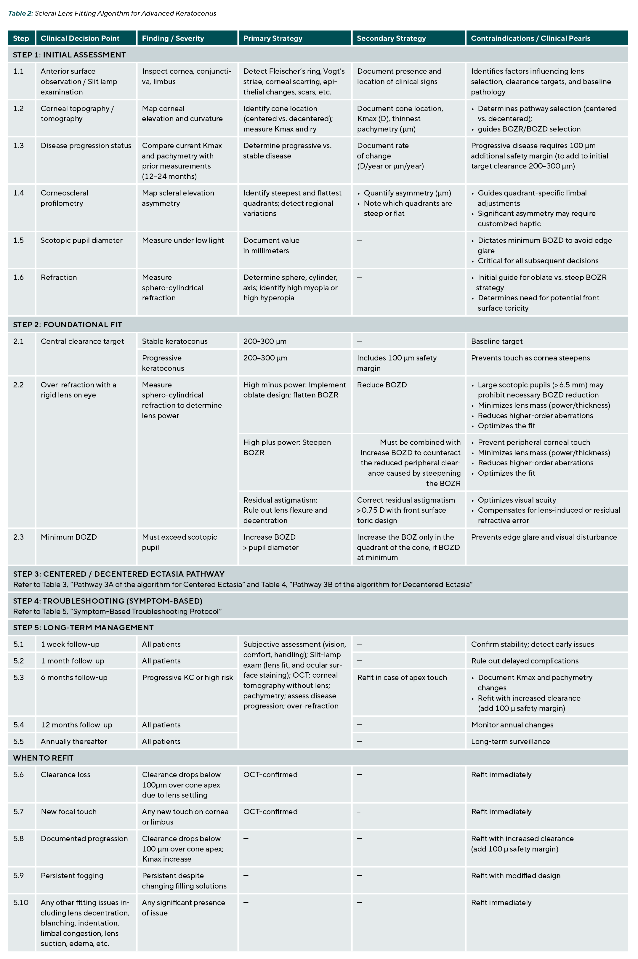
Keratoconus is a progressive disease. Even after corneal cross-linking, some eyes continue to steepen at a slow rate.32 A scleral lens fitted with low apical clearance (< 200 μm) at dispense may develop focal corneal touch within a year as the cone elevates, leading to scarring and reduced visual acuity.22,33 Conversely, excessive clearance (> 400 μm) may reduce oxygen delivery and cause different issues, including lens decentration and midday fogging.31 The optimal balance is a target post-settling central clearance of 200–300 microns for stable keratoconus, with an additional ~100-micron safety margin for documented progressive keratoconus.
Patients fitted with the 100-micron progression safety margin require follow-up every six months to monitor disease progression and confirm no settling-induced touch. If the lens is touching the apex despite the initial safety margin, it should be replaced with one that offers greater vault.
Pillar 2: Manipulation of BOZR and BOZD to Reduce Lens Power and Optimize Optical Performance
Foundational Concept of Master Parameter Control: BOZR and BOZD Dynamics
While BOZR and BOZD are well recognized as crucial parameters in corneal lens fitting, their importance is often overlooked in scleral lens practice, where clinicians tend to focus primarily on lens sagittal values, elevation, and haptic alignment on the conjunctiva. However, successful scleral lens fitting also rests on understanding these two primary parameters and their reciprocal relationship. When clinicians struggle to adjust the haptic lens to resolve alignment issues, the root cause often lies in unaddressed problems at the corneal periphery and limbus. If these corneal peripheral and limbal zones are not properly managed, alignment issues will persist, manifesting as limbal congestion, conjunctival blanching, impingement, midday fogging, lens suction, conjunctival prolapse, lens decentration, and persistent discomfort.29,30,31,34 Unlike empirical trial-and-error, a parameter-driven approach that deliberately manipulates BOZR and BOZD enables the clinician to predictably achieve the desired fit and optical outcomes in the peripheral cornea and over the limbus.
Manipulating BOZR and BOZD: Impact on Lens Fit
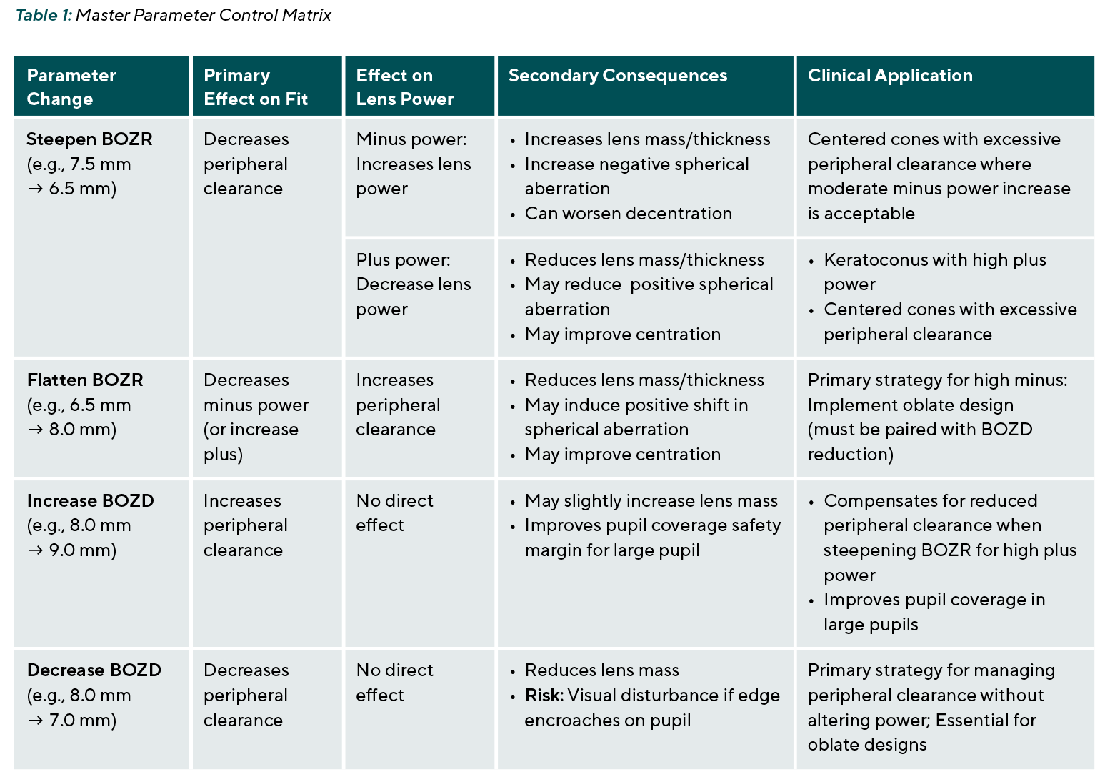
The interaction between BOZR and BOZD follows several predictable patterns, and they are summarized in Table 1. Steepening the BOZR (e.g., from 7.5 mm to 6.5 mm) decreases peripheral clearance and increases minus power. This maneuver is useful for centered cones with excessive peripheral clearance, where a moderate increase in power is acceptable, though it increases lens mass and may worsen decentration. Flattening the BOZR (e.g., from 6.5 mm to 8.0 mm) decreases minus power while increasing peripheral clearance. However, flattening the BOZR always requires reducing the BOZD to compensate for the newly created excessive peripheral clearance. This is the core strategy for high myopia
(oblate design).
Increasing the BOZD (e.g., from 8.0 mm to 9.0 mm) increases peripheral clearance without a direct effect on power. This adjustment compensates for reduced clearance when steepening the BOZR to reduce high-plus power. Decreasing the BOZD (e.g., from 8.0 mm to 7.0 mm) reduces peripheral clearance without altering power. This is the primary strategy for managing excessive peripheral clearance in centered cones and is essential for controlling clearance in oblate designs.
The High Myopia Paradox in Conventional Scleral Lens Fitting
In advanced keratoconus complicated by high myopia, conventional scleral lens fitting presents a significant clinical paradox. To achieve an adequate central vault on a steep cornea, the practitioner must select a relatively steep BOZR.35 The optical consequence of this geometry is the generation of high minus power, often reaching –15.00 diopters or more. Physically, the lens becomes excessively thick at its center, which adds considerable mass. This heavy, thick lens tends to decenter more easily, reducing comfort and stability. Moreover, the high-minus power induces significant image minification, typically reducing the retinal image size and directly degrading both high- and low-contrast visual acuity.35
Oblate Geometry: Flipping the Paradigm
The oblate design offers a fundamental rethinking of scleral lens geometry for the highly myopic keratoconic eye. Unlike the prolate lens, which is steeper centrally and flatter peripherally, an oblate lens has a flatter central curve than its peripheral curves. By deliberately flattening the BOZR – for example, from 6.5 mm to 8.0 mm – the practitioner can maintain central vault not through central steepness but by strategically steepening the peripheral transition zone, which effectively „pinches“ the lens, generating the necessary sagittal height to clear the cone apex without relying on a steep central curve. The flatter BOZR directly reduces the required minus power, which dramatically lowers lens mass, center thickness, and image minification. A reduction in BOZD is needed to reduce the peripheral clearance created by the flatter BOZR. Before implementing this approach, a scotopic pupil measurement is essential to determine the absolute minimum BOZD to avoid visual disturbance caused by encroachment of the optic zone edge. The oblate design thus transforms a problematic high minus lens into a more physiologically and optically tolerable device.
Managing High Plus Power: A Separate Challenge
Although less common, certain cases of keratoconus may require high plus power.36 While high myopia has received considerable attention, high plus power presents an equally important but distinct challenge in scleral lens fitting for keratoconus.
In conventional scleral lens fitting for high plus power, a relatively flat BOZR is typically required to achieve the desired positive power. However, a flat BOZR increases peripheral clearance.
The solution for high plus power is to steepen the BOZR, which directly reduces positive power. This reduction in plus power has several important benefits: it decreases the lens center thickness, mass, and decentration. However, steepening the BOZR also decreases peripheral clearance, which can lead to peripheral corneal touch and limbal touch. Therefore, any significant steepening of the BOZR for high plus power may need to be accompanied by an increase in BOZD to counteract the reduction in peripheral clearance. This combination – steeper BOZR plus larger BOZD – maintains a safe peripheral vault while reducing the required plus power, resulting in decreased lens thickness and mass, better centration, and greater comfort.
Contraindications for Power-Adjusting Strategies
Despite their advantages, both oblate (high-minus) and steepened BOZR (high-plus) strategies have limitations. For high minus, the most important contraindication is a large scotopic pupil diameter exceeding 6.5 mm. Because the oblate design relies on a reduced BOZD, the optic zone edge may encroach on the pupil under low-light conditions, causing significant edge glare, halos, or other disturbing visual phenomena.29,34 In such patients, a conventional steep BOZR with a larger BOZD remains the preferred approach, accepting the higher minus power and its associated minification as a necessary trade-off to avoid disabling
night-vision symptoms.
For high plus power, the main limitation is that steepening the BOZR may not be feasible in eyes with extremely steep corneas, where the required BOZR would be impractically small. In these cases, alternative strategies include accepting higher plus power with a flatter BOZR and larger BOZD.
Pillar 3: Management of Scleral Asymmetry
Research using corneoscleral topography has revealed several key patterns in keratoconic eyes. There is a positive correlation between corneal irregularity and scleral asymmetry; in essence, a more irregular cornea predicts a more irregular sclera.37 A study has shown that the keratoconic eye has a much more asymmetrical corneoscleral profile than the healthy eye, especially in moderate-to-advanced disease.38 The report also found that scleral steepening tends to mirror corneal steepening.38 Similarly, another study reported significant differences in sagittal height variation across meridians between ectatic and normal eyes.39 A strong correlation emerged between the magnitude of cone decentration and the level of scleral asymmetry, indicating that greater cone displacement predicts more pronounced scleral contour abnormalities in advanced disease.39
This relationship in earlier-stage keratoconus, where cone decentration is less pronounced, was later explored.37 It has been found that a moderate correlation exists between cone decentration and scleral toricity, suggesting that disease severity affects the extent to which cone position predicts scleral shape. The study also showed that when a corneal meridian steepens, the corresponding scleral semi-meridian steepens as well.37 However, the reverse did not hold – corneal flattening did not predict scleral flattening. Additionally, the angle between the steepest and flattest regions of the sclera varies significantly from patient to patient, meaning that corneal measurements alone cannot reliably determine the optimal toric lens design.37
Another report examined whether habitual lens wear alters corneoscleral shape by comparing three severity-matched keratoconus groups (lens-naive, corneal lens wearers, and scleral lens wearers) against healthy emmetropic controls.40 The findings showed that both types of contact lens wear induced localized shape changes in the peripheral cornea and sclera, and keratoconic eyes had significantly steeper peripheral corneas and scleras than healthy eyes. Among the keratoconus groups, scleral lens wearers exhibited the highest degree of scleral asymmetry.40 This suggests that a scleral lens with a spherical landing zone will likely result in uneven tissue compression across quadrants in these patients.
Bottom Line
Several consistent patterns emerge from this body of literature.
• First, keratoconic eyes demonstrate a highly asymmetric corneoscleral profile compared to healthy eyes.
• Second, as keratoconus progresses, the sclera not only becomes steeper but also more irregular, particularly in the same semi-meridians where corneal steepening occurs.
• Third, the peripheral cornea and sclera are significantly steeper in keratoconus than in normal eyes.
• Fourth, corneal topographic data can offer useful clues about the presence and location of scleral asymmetry, especially the steepest scleral angle.
• Fifth, in moderate-to-advanced disease, the degree of cone decentration may serve as a clinical indicator of underlying scleral asymmetry.
Clinical Implications for Scleral Lens Fitting
These anatomical insights carry direct practical consequences for the scleral lens practitioner. The presence of cone decentration should raise suspicion of concomitant scleral asymmetry and prompt consideration of a quadrant-specific or customized landing zone design, rather than relying on a symmetric spherical or toric haptic. Furthermore, when fitting scleral lenses in advanced keratoconus, the lens periphery may need to be steeper than conventional fitting norms would suggest to align with the naturally steeper scleral contour characteristic of this disease. Failing to account for these regional variations risks poor lens centration, focal conjunctival compression, reduced wearing time, and suboptimal visual outcomes.31
Comprehensive Clinical Algorithm: Centered Versus Decentered Ectasia
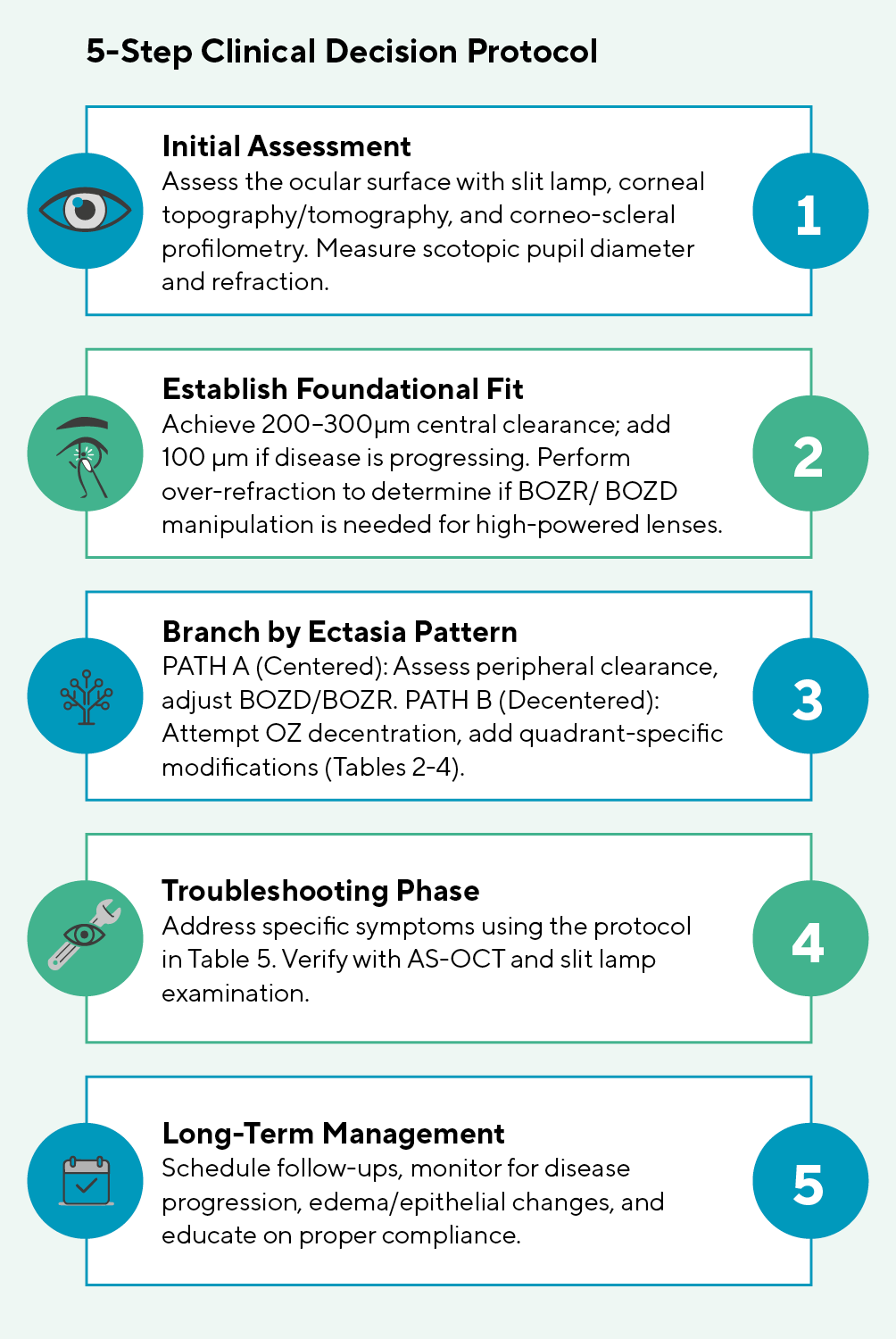
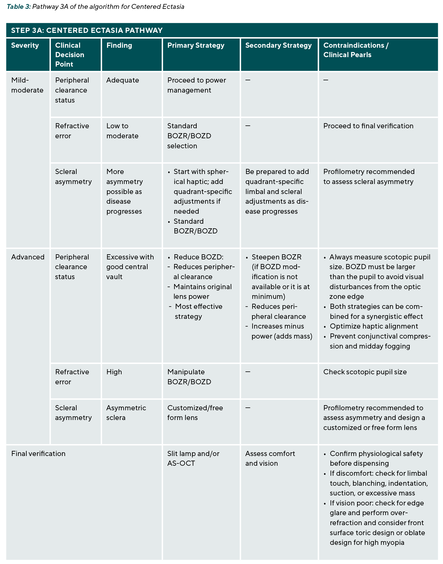
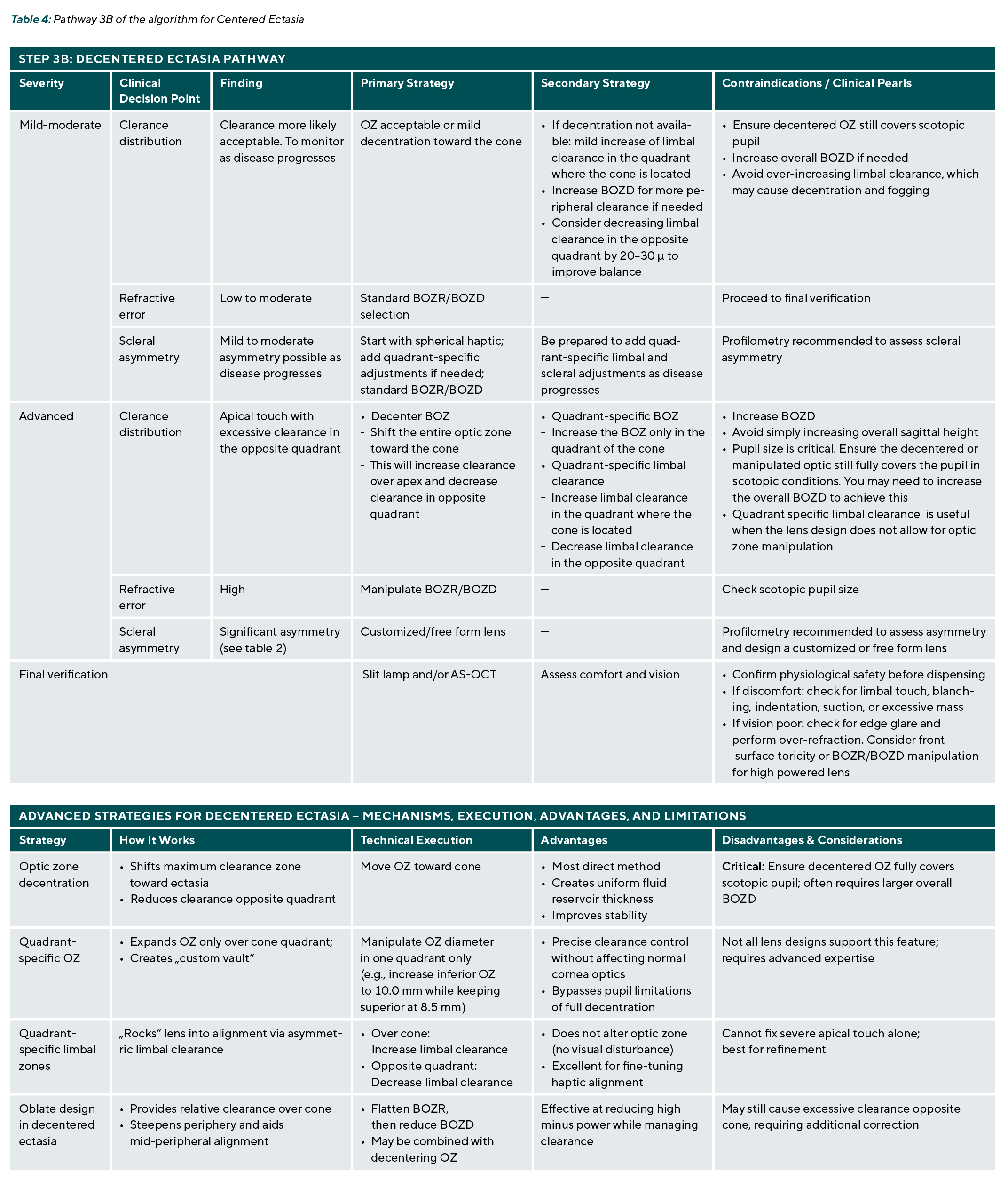
The following stepwise algorithm synthesizes the key principles outlined in the preceding sections into a practical, clinical decision tree for fitting scleral lenses in advanced keratoconus. The algorithm begins with essential pre-fitting assessments, including slit lamp examination to detect clinical signs such as Fleischer‘s ring, Vogt‘s striae, corneal scarring, and epithelial changes that may influence lens selection and clearance targets, and corneal topography and tomography to identify cone location, maximum keratometry (Kmax), thinnest pachymetry, and documentation of disease progression over the preceding 12 to 24 months. Scotopic pupil diameter must be measured, as it determines the minimum acceptable BOZD to avoid edge glare. Corneoscleral profilometry is also crucial for assessing scleral asymmetry, reducing chair time and lens reorders. A manifest refraction is necessary to estimate lens power and determine whether high myopia or high hyperopia is present, guiding the choice between flat and steep BOZR strategies. Based on these data, the practitioner first achieves a foundational fit with appropriate central clearance and establishes the 100 micron progression safety margin. A rigid lens is then applied, and an over refraction is performed to determine whether manipulation of the BOZR or BOZD is required for high-powered lenses. The fitting then branches into distinct pathways for centered versus decentered ectasia. The algorithm concludes by troubleshooting common complications and outlining long-term follow-up, and is presented in Tables 2–4 and Figure 1.
Pathway A: Centered Ectasia
In patients with a centered cone, the fitting strategy is guided by disease severity (mild-moderate vs. advanced), peripheral clearance status, refractive error, and the presence or absence of scleral asymmetry.
For mild to moderate disease, peripheral clearance is typically adequate, allowing the practitioner to proceed directly to power management. Refractive error is usually low to moderate, and standard BOZR and BOZD selection is sufficient. However, as the disease progresses, scleral asymmetry may develop. In such cases, the practitioner should start with a spherical haptic and standard BOZR and BOZD but remain prepared to add quadrant-specific limbal and scleral adjustments as needed. Corneoscleral profilometry is recommended to rule out scleral asymmetry.
In advanced-centered ectasia, excessive peripheral clearance with good central vault is a common finding. The first-line strategy is to reduce the BOZD, which decreases peripheral clearance while maintaining the original lens power. If the BOZD modification is not available or the BOZD is already at its minimum safe limit, an alternative is to steepen the BOZR, which also reduces peripheral clearance but increases minus power and adds lens mass. Both strategies can be combined for a synergistic effect. Regardless of the approach, the practitioner must always verify that the BOZD remains larger than the scotopic pupil to avoid visual disturbances from the optic zone edge. The goals of this step are to optimize haptic alignment, prevent conjunctival compression, and reduce the risk of midday fogging. When significant scleral asymmetry is present in advanced disease, a customized or free-form lens design is recommended, guided by corneoscleral profilometry.
Pathway B: Decentered Ectasia
In patients with a decentered cone, the fitting strategy differs fundamentally from centered ectasia, as symmetric lens designs invariably lead to low or apical touch on the cone and excessive vault in the opposite quadrant. As with centered ectasia, the approach is guided by disease severity (mild-moderate versus advanced), distribution of clearance, refractive error, and the degree of scleral asymmetry.
For mild to moderate disease, a clearance distribution is more likely to be acceptable, but the patient should be monitored as the disease progresses. The primary strategy is to use an optic zone that is either acceptable as is or with mild decentration toward the cone. If decentration is not available, a mild increase of limbal clearance in the quadrant where the cone is located can be effective. Additionally, increasing the BOZD may provide more peripheral clearance if needed, and decreasing limbal clearance in the opposite quadrant can uniform the vault. The practitioner must ensure that the decentered OZ still fully covers the scotopic pupil. It is crucial to avoid over-increasing limbal clearance, which may cause decentration and fogging. For patients with low to moderate refractive error, standard BOZR and BOZD selection is appropriate. When mild to moderate scleral asymmetry is present, the practitioner should start with a spherical haptic and standard BOZR and BOZD, but remain prepared to add quadrant-specific limbal and scleral adjustments as the disease progresses. Corneoscleral profilometry is recommended to rule out subclinical asymmetry.
In advanced decentered ectasia, clearance distribution is characterized by low apical clearance or apical touch with excessive clearance in the opposite quadrant. The primary strategy is to decenter the back optic zone, shifting the entire optic zone toward the cone, which increases clearance over the apex and decreases clearance in the opposite quadrant. If the lens design does not support optic zone decentration, alternative strategies include quadrant-specific BOZ modification (increasing the BOZD only in the quadrant of the cone) or quadrant-specific limbal clearance adjustments (increasing limbal clearance in the cone quadrant and decreasing it in the opposite quadrant). The practitioner may increase the overall BOZD as needed and avoid simply increasing the overall sagittal height, as this will exacerbate the vault opposite the cone. Pupil size is critical; the decentered or manipulated optic must still fully cover the pupil under scotopic conditions, which may require increasing the overall BOZD. Quadrant-specific limbal clearance is particularly useful when the lens design does not allow for optic zone manipulation. When significant scleral asymmetry is present in advanced disease, a customized or free-form lens designed with corneoscleral profilometry is recommended. In summary, advanced strategies for managing decentered ectasia include optic zone decentration, quadrant-specific optic zone expansion, quadrant-specific limbal curve adjustments, and the oblate design combined with decentration, each with specific advantages and limitations as summarized in the corresponding table.
Final Verification (Common to Both Pathways)
Regardless of whether the ectasia pattern is centered or decentered, final verification follows the same principles. Slit lamp examination and anterior segment OCT are performed to confirm physiological safety before dispensing. The clinician must verify that central clearance is within the target range, that no focal touch is present, that peripheral clearance is uniform, and that the lens is more likely well-centered. If the patient reports discomfort, the clinician should check for limbal touch, conjunctival blanching, indentation, suction, or excessive lens mass. If vision is poor, the clinician should evaluate for edge glare, perform an over-refraction, and consider a front-surface toric design for residual astigmatism exceeding 0.75 D, or manipulate BOZR/BOZD for high-powered lenses. This systematic verification ensures that both centered and decentered ectasia cases achieve optimal visual function and long-term ocular health before the lens is dispensed.
The Evolving Role of Piggyback Systems
A piggyback system in the context of scleral lenses refers to the use of a soft contact lens placed either under the scleral lens (between the scleral lens and the cornea/sclera) or over the scleral lens (on its front surface). While traditionally associated with corneal lenses, piggyback configurations have specific, evidence-based applications in scleral lens wearers with complex ocular surface conditions.29,34,41,42,43,44
In progressive keratoconus, the cornea may continue to steepen over time. A scleral lens that provides adequate clearance at the initial fitting may develop apical touch within 6 to 12 months. If the lens warranty has expired and the patient cannot afford a replacement, placing a soft contact lens beneath the scleral lens will act as a cushion, preventing touch and mechanical stress on the cone.29,34 This approach is especially useful in pediatric or rapidly progressive keratoconus, where frequent lens replacements are burdensome. However, it is only a temporary solution intended to bridge the gap until a new lens can be obtained, typically within two years, although replacement intervals may vary depending on several factors, including but not limited to lens condition and clinical needs.45
A limitation of the piggyback system is the potential for reduced oxygen supply to the cornea; however, a short-term study in healthy young adults found that a reverse piggyback scleral lens did not induce clinically or statistically greater central corneal edema compared to standard scleral lens wear.46
Troubleshooting Common Complications
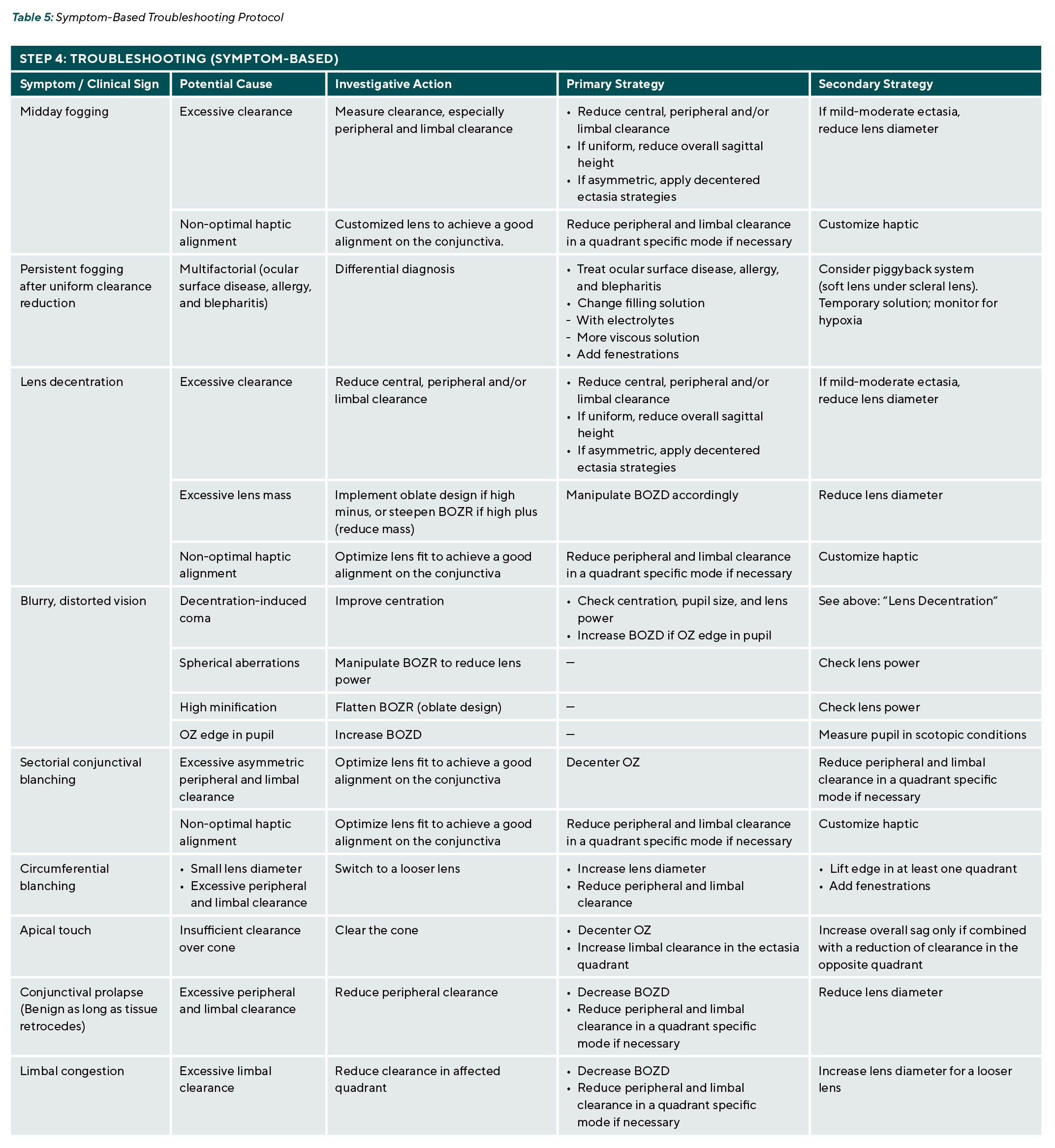
Despite careful initial fitting, patients with advanced keratoconus may present with specific symptoms or clinical signs that indicate underlying lens design or fit issues. Rather than resorting to trial-and-error adjustments, a systematic approach guided by clinical observation allows the practitioner to identify the root cause and implement targeted parameter modifications. Table 5 summarizes the most common complications encountered in scleral lens wearers with keratoconus, their potential causes, recommended investigative actions, and the corresponding parameter adjustments to resolve each issue.
Integration of All Three Pillars
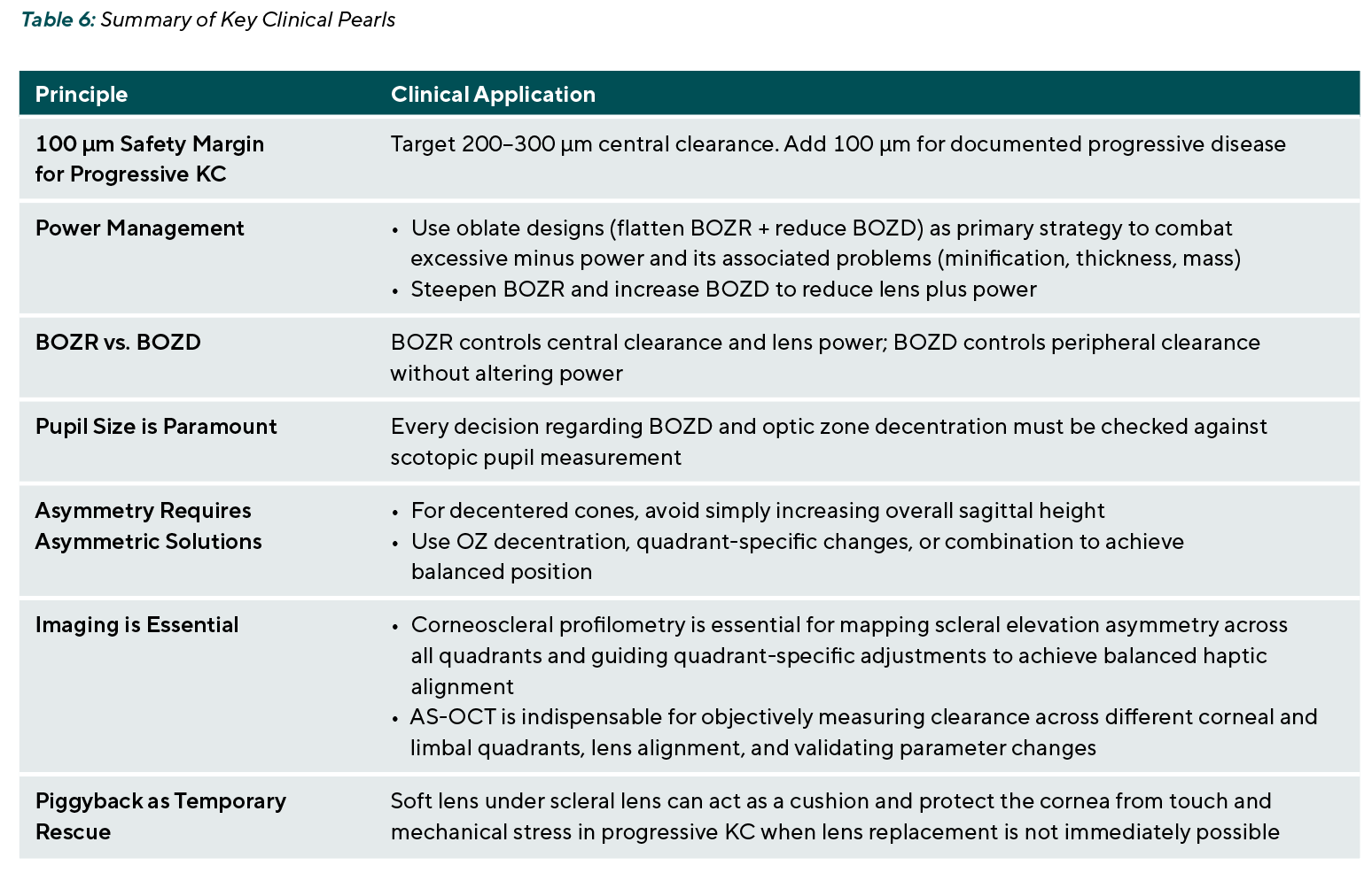
The three pillars described in this article – the 100-micron progression safety margin, the manipulation of BOZR and BOZD, and the scleral asymmetry, alongside the comprehensive algorithm for centered versus decentered ectasia and the piggyback system – are not independent options but rather integrated components of a single, unified fitting strategy. For any given patient, the practitioner begins with the pre fitting data collection, then selects the appropriate ectasia pathway (centered or decentered), applies the progression safety margin, addresses high-power lenses if indicated, and reserves piggyback configurations. Clinical observations serve as the common thread, validating each step and guiding iterative adjustments. By following this structured, evidence based approach, even the most challenging advanced keratoconus cases can be successfully rehabilitated with scleral lenses, achieving the dual goals of optimal visual function and long term ocular health. This algorithm assumes the availability of fully customizable scleral lens designs. Economic and access disparities may limit implementation. Table 6 summarizes the key clinical pearls.
Conclusion
Advanced keratoconus management with scleral lenses requires a systematic, parameter-driven approach. By understanding the distinct effects of BOZR and BOZD manipulations and by applying targeted strategies based on ectasia pattern and scleral asymmetry, practitioners can achieve successful, reproducible outcomes. The presented algorithm offers a structured clinical pathway that integrates current evidence with practical fitting principles, advancing the standard of care for this complex patient population.
Conflict of interest
The author is a consultant for Wave Eye Care and has received financial support or lecture honoraria from the following companies: Bausch & Lomb, Boston Materials, Contamac, Eaglet Eye, EYEXY, Medlac, Mediworks, Medmont, Multilens, 3N Eyecare, Oculus, Topcon Healthcare, Wave Eye Care.
Jedlicka, J., Farajian, T. S., van der Worp, E. (2019). Correlation of Corneal and Scleral Topography in Cases with Ectasias and Normal Corneas. J. Cont. Lens Res. and Sci., 3:e10-e20.